
Radiology in Medical Diagnosis
Imaging methods serve as the “eyes” of modern medicine. It is impossible to properly treat a disease that has not been diagnosed. For this reason, radiology is not merely a department that takes X-rays; it is the most critical branch of medicine, one that alters the entire course of clinical decisions and determines the direction of treatment.
Radiological imaging methods involve mapping the body’s internal structures using X-rays (radiation), sound waves, or strong magnetic fields. Each test has its own unique physical operating principle, advantages, and limitations. The “most advanced” device for a given disease may not always be the “most accurate” one. To arrive at the correct diagnosis, it is essential to use the right device to answer the right clinical question. Below, you will find the most commonly used radiological methods in medicine, along with the fundamental medical facts to keep in mind.
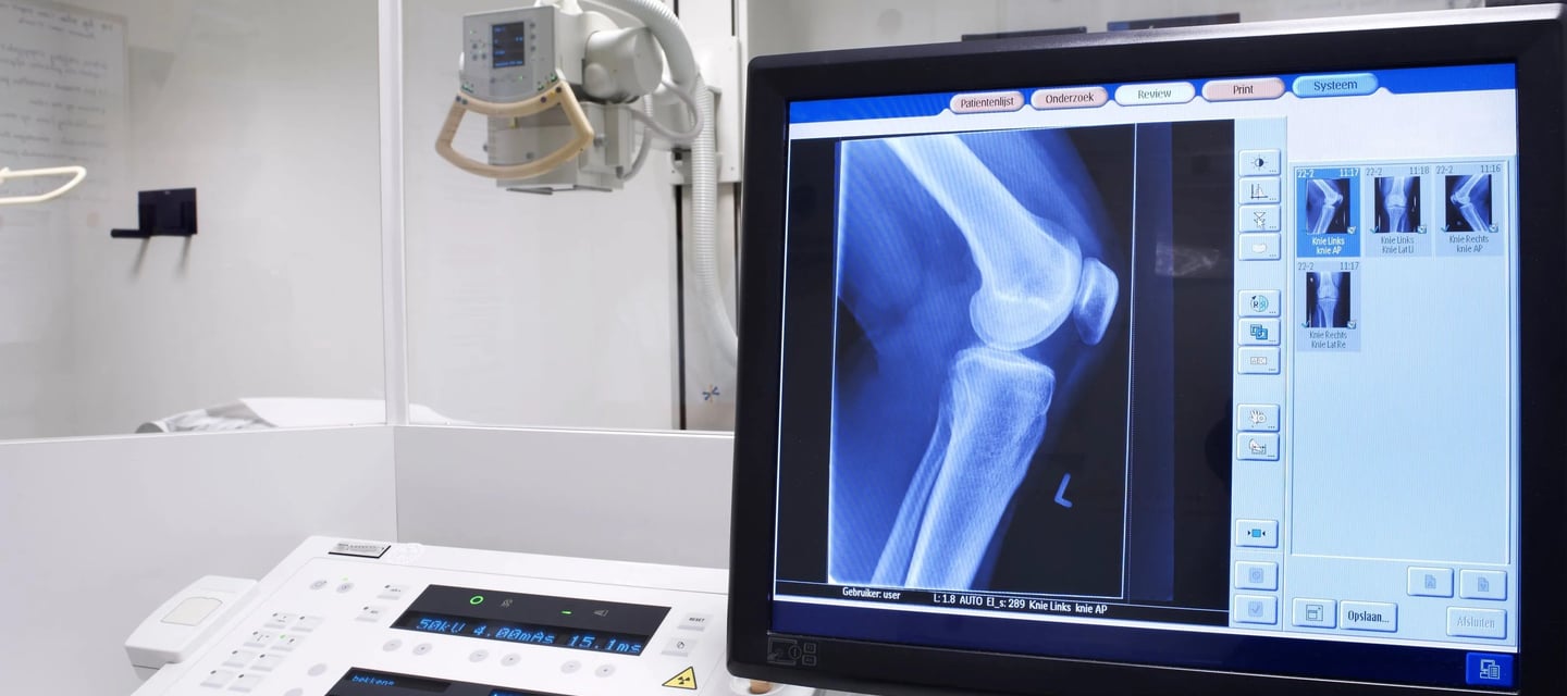
Imaging Methods and Principles of Operation
Direct imaging (X-ray): It is one of the oldest and fastest imaging methods. Using a low dose of X-rays (radiation), it examines the integrity of bone structures, the lung parenchyma, and the condition of the sinuses. It is indispensable for the initial diagnosis of emergencies such as bone fractures, lung infections (pneumonia), and intestinal blockages (ileus).
Computed Tomography (CT): It uses X-rays that rotate around the body to produce cross-sectional and 3-dimensional images. It provides much greater detail than a standard X-ray. It is indispensable in emergency departments, particularly for the rapid detection of brain hemorrhages following head trauma, pulmonary embolism, and injuries to internal organs. However, because the radiation dose is much higher than that of a standard X-ray, it should be used only when absolutely medically necessary.
Ultrasonography (USG) and Doppler: It is a completely safe, radiation-free method that uses sound waves (ultrasound). A real-time image is created as sound waves reflect off tissues. It is the first-choice method for monitoring abdominal organs (liver, gallbladder, kidneys), the thyroid gland, breast tissue, and the fetus during pregnancy. Doppler ultrasound is used to examine the speed and direction of blood flow in blood vessels to detect clots or narrowing.
Magnetic Resonance Imaging (MRI): Images are obtained by detecting the vibrations of hydrogen atoms in response to a very strong magnetic field and radio waves. It does not involve radiation. It is the gold standard for visualizing soft tissues such as the brain, spinal cord, nerves, muscles, ligaments, and joints. It is the most accurate method for determining the extent of tissue loss in cases of hernias, meniscus tears, brain tumors, and stroke.
Clinical Warnings and Safety Protocols
Contrast Agents and Kidney Health: During CT and MRI scans, a contrast agent (commonly referred to as “contrast” or “contrast-enhanced imaging”) is injected into a vein to make blood vessels or tissue boundaries more visible. These substances are excreted from the body through the kidneys. For this reason, in patients with kidney failure or those at risk, a creatinine test must be performed to check kidney function before administering a contrast agent. Otherwise, acute kidney damage may be triggered.
MRI Machines and Metal Safety: An MRI machine is a massive, constantly active magnet. Patients with pacemakers (older, non-MRI-compatible models), cochlear implants, or metal shrapnel or prostheses in their bodies must not enter the MRI room under any circumstances. These metals can shift due to the machine’s powerful magnetic field, causing them to heat up and potentially resulting in tissue burns or fatal internal injuries.
Radiation Sensitivity (Children and Pregnant Women): Children’s cells divide at a very high rate; their tissues are many times more sensitive to radiation than those of adults. For this reason, in children and pregnant women (except in cases of absolute necessity), ultrasound (USG), which uses sound waves, or magnetic resonance imaging (MRI), which uses magnetic fields, should be preferred over CT scans and X-rays, which involve ionizing radiation.
Critical Error Warning: The idea that “I’ll pay more so we can get the most expensive MRI right away” is completely incorrect from a medical standpoint. For example, to detect a gallstone or sludge in the gallbladder, a dynamic upper abdominal ultrasound—rather than an MRI or CT scan—provides much more sensitive and accurate results. Your clinician should decide which test to perform.
When should advanced imaging be performed?
After a physical examination, your doctor will order the necessary tests based on your clinical presentation. However, in the following situations, an urgent and rapid radiological evaluation is of vital importance:
Severe head trauma, a fall from a height, or a traffic accident (requires an urgent CT scan)
Sudden swelling, redness, increased warmth, and severe pain in the leg (Doppler ultrasound for suspected deep vein thrombosis)
Severe abdominal pain localized to the right lower quadrant, accompanied by fever and vomiting (Ultrasound or CT scan for suspected appendicitis)
Shortness of breath, coughing up blood, or sudden chest pain (CT angiography for suspected pulmonary embolism)
Sudden weakness on one side of the body, speech or vision problems (contrast-enhanced brain MRI for acute stroke evaluation)
Important Note: Radiology reports are not the work of the technician who takes the images; rather, they are the specialized work of radiologists who interpret the thousands of shades of gray, anatomical abnormalities, and pathologies visible in the images. The quality of your tests and their accurate interpretation are the most fundamental prerequisites for the success of your treatment. Please contact our clinic for any concerns regarding your imaging procedures or to schedule an appointment.
Merve Medical Center
Reliable and accessible healthcare in Istanbul since 1992..
CHAPTERS
Home
Our Units
About
Contact
CONTACT
info@mervetipmerkezi.com
+90 212 537 77 79
İstanbul, Türkiye
7/24 Emergency & Outpatient Services
© 2026 Merve Medical Center - Uninterrupted medical care in Istanbul since 1992.
Editor: Dr. Gökhan Çetinkaya Web Design: Nahit Demir Last Updated: September 26, 2026
CLINICAL EXPERTISE AND TRUST